Fig 2.
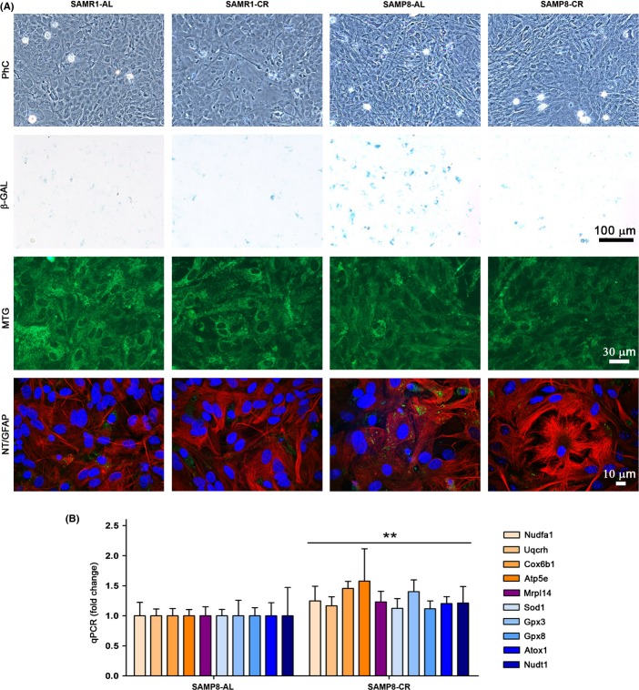
Senescence phenotype of SAMP8 astrocytes was ameliorated by caloric restriction. (A) No distinctive morphological changes were observed by phase contrast microscopy (PhC) in SAMP8 as compared to control strain SAMR1, but β-galactosidase (β-GAL) blue staining of the same microscopy fields indicated the presence of replicative senescence in SAMP8 astrocytes treated with ad libitum serum (SAMP8-AL), whereas staining was greatly reduced in those treated with caloric restriction serum (SAMP8-CR). Mitotracker green FM (MTG) stained a similar mitochondrial pattern in the different cultures (green fluorescence). Immunostaining of nitrotyrosinated proteins (NT) was only visible (green fluorescence spots) in SAMP8-AL astrocytes, whereas that of GFAP (red fluorescence) showed similar astrocyte morphology in the different cultures; nuclei were counterstained with TO-PRO-3 iodide (confocal images). Representative images, n = 4. (B) Gene expression of mitochondrial and antioxidant genes measured by real-time qPCR showed a moderate increase in SAMP8 astrocytes after CR treatment. Nudfa1, Uqcrh, Cox6b1, Apt5e, and Mrpl14 encode proteins of mitochondrial complexes I, III, IV, V, and mitochondrial ribosomes, respectively; Sod1, Gpx3, Gpx8, and Atox1 encode a variety of soluble and membrane antioxidants, and Nudt1 a selective nucleic acid antioxidant. See Table S7 (Supporting information) for full name of genes and oligomers used. Statistics: **P < 0.01 overall effect of CR treatment according to ANOVA, without post hoc Bonferroni's test significance.

