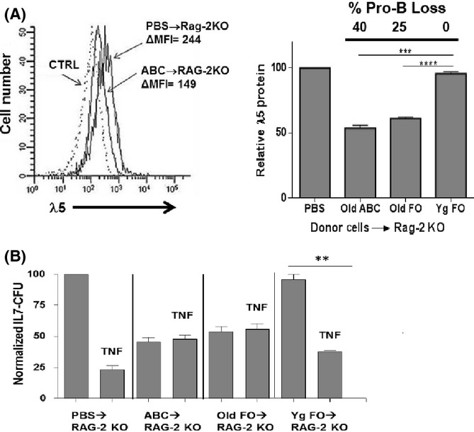
Fig 5.

Residual pro-B cells that remain in RAG-2 KO bone marrow after adoptive transfer of old B cells are low in surrogate light chain λ5 and resist TNFα-induced apoptosis. (A, left) Representative analysis of cytoplasmic λ5 protein, detected by fluorescent staining with LM34 mAb, in pro-B cells from B6 RAG-2 KO mice either untreated (PBS injection only) or 4 weeks after adoptive transfer of 1.7 × 10⁶ splenic ABC, pooled from 3 old B6 mice (22–31 months of age). (A, right) Cumulative data is shown on pro-B-cell loss and λ5 protein expression for B6 RAG-2 KO recipients of either PBS, old B6 splenic ABC, or young or old B6 splenic follicular B cells (four mice per group) 1 month after adoptive transfer. Pro-B cell loss in the recipient bone marrow is also indicated for each group. Relative pro-B λ5 protein was calculated as the λ5 Δmean fluorescence intensity (MFI) for each group divided by the λ5 ΔMFI for the pro-B cells in PBS-treated control RAG-2 KO mice (100%). (B) Bone marrow cells from the adoptively transferred mice in (A) were cultured (1 × 10⁶ ml−1) in the presence/absence of optimal concentrations (10 ng ml−1) of rmTNFα in IL-7 CFU assays. Normalized IL-7 CFU for each experimental group were reported relative to the IL-7 CFU numbers for the control group (100%). Statistical analysis (two-tailed Student's t-test): **P < 0.05; ***P < 0.01; ****P < 0.001.
