
Fig 3.
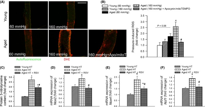
Aging exacerbates hypertension-induced vascular oxidative stress. A: Representative confocal images showing stronger dihydroethidium (DHE) staining (red fluorescence) indicating increased O2.− production in high pressure-exposed MCAs isolated from aged mice as compared to MCAs isolated from young mice. MCAs were pressurized at 60 and 160 mmHg for 6 h. Note that high pressure-induced oxidative stress in aged MCAs was significantly attenuated by the NADPH oxidase inhibitor apocynin and the mitochondria-targeted antioxidant mitoTEMPO. Green autofluorescence is shown for orientation purposes (original magnification: 20×, scale bar: 100 μm). Bar graphs (B) are summary data. Data are means ± SEM (n = 6 in each group). *P < 0.05. vs. Young (160 mmHg),#P < 0.05 vs. Aged (160 mmHg). C: Cortical 3-nitrotyrosine content in hypertensive (HT) young, aged, and resveratrol-treated aged mice (n = 6 in each group). Data are mean ± SEM. *P < 0.05 vs. Young HT, #P < 0.05 vs. Aged HT. D, E, and F show hypertension-induced mRNA expression of NADPH oxidase subunits Nox2, Nox4, and Ncf1 in cerebral arteries of young, aged, and resveratrol-treated aged mice. Data are mean ± SEM (n = 6 in each group). *P < 0.05 vs. Young HT, #P < 0.05 vs. Aged HT.