
Fig 2.
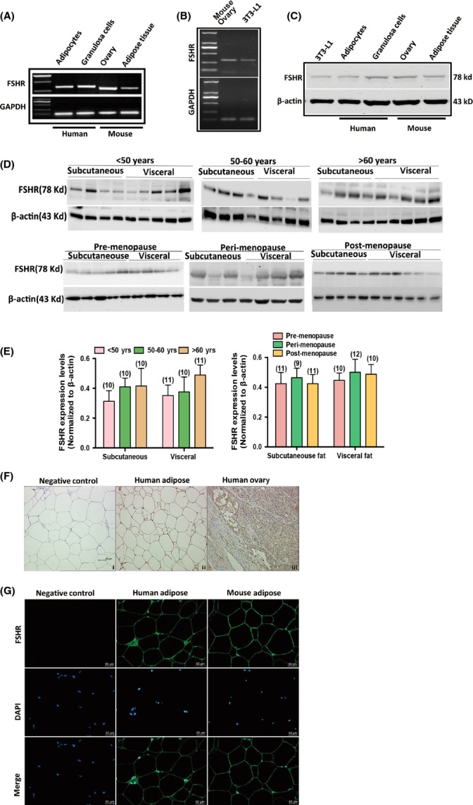
Expression and localization of FSHR in human and mouse adipocytes. (A) mRNA expressions of FSHR in adipocytes and granulosa cells of human and in adipose tissue and ovarian tissue of mouse. GAPDH served as loading control. (B) mRNA expression of FSHR in 3T3-L1 preadipocytes. GAPDH served as loading control. (C) Protein expressions of FSHR in 3T3-L1 preadipocytes, adipocytes and granulosa cells of human and in adipose tissues and ovarian tissues of mouse. β-actin served as loading control. (D) Protein expression of FSHR in subcutaneous and visceral fat of males aged < 50, 50–60 and > 60 years and in pre-, peri- and postmenopausal females. (E) Relative protein levels of FSHR in subcutaneous and visceral fat of pre-, peri- and postmenopausal females. Values are mean ± SEM; the number of samples in each group is shown at the bottom of the column. No significant differences were observed between groups. (F) Localization of FSHR in human adipose and ovarian tissues by immunohistochemistry. (G) Localization of FSHR in human and mouse adipose tissues by immunofluorescence. Nuclei were stained with DAPI.