Fig 1.

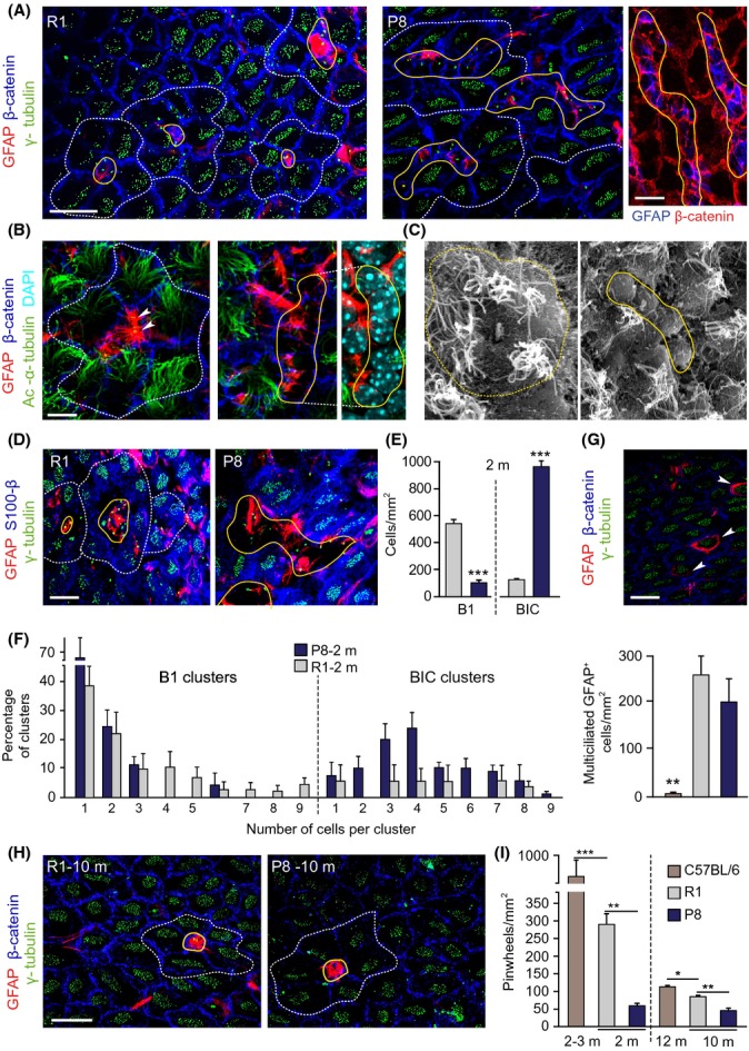
The cytoarchitecture of the SEZ is altered in young P8 mice. (A) Whole-mounts from 2-m SAMR1 (R1) and SAMP8 (P8) mice immunostained for GFAP, β-catenin, and γ-tubulin. (B) GFAP, β-catenin, and acetylated α-tubulin. Arrowheads point to B1-cell primary cilium. (C) Whole-mounts in scanning electron microscopy. Note the presence of misshapen pinwheels with abutted GFAP cells in P8 mice. (D) GFAP, γ-tubulin, and S100β in R1 (left) and P8 (right) whole-mounts. BICs are S100β−. (E) Number of B1 cells and BICs in SEZs at 2-m. (F) Distribution of GFAP+ B1 cells (left) or BICs (right) at 2-m. (G) GFAP, γ-tubulin, and S100β in whole-mounts showing GFAP+ multiciliated cells (white arrowheads) in the plane of the ependyma (above) and quantification of these cells (below) in 2-m R1, P8, and C57BL/6 mice. (H) GFAP, γ-tubulin, and β-catenin in SEZ whole-mounts at 10-m. Only normally organized pinwheels are observed in both genotypes. (I) Pinwheel density in the dorsal SEZ of 2- and 10-m R1 and P8 mice, and in 3- and 12-m C57BL/6 mice. Pinwheels are delineated by dashed white lines, and B1 cells/BICs are delineated by solid yellow lines. Data are shown as mean ± SEM of the indicated number of mice (n) from each strain (*P < 0.05; **P < 0.01; ***P < 0.001). Scale bars: (A, left) 10 μm; (B) 5 μm; in (A, right), (D), (G) and (H) 20 μm.
