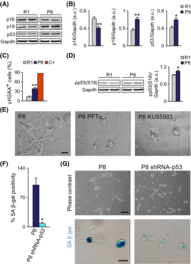
Fig 4.

Senescence of P8 cells requires p53. (A) Left: Representative immunoblots for p16, p19, and p53 in P2 neurospheres from 2-m R1 and P8 mice. (B) Densitometric quantification of p16, p19, and p53 relative to Gapdh levels (R1 n = 7, P8 n = 7). (C) Percentage of cells with γ-H2AX+ foci. Positive control (C+) is a doxorubicin-treated (0.5 μg/ml, 6 h) neurosphere culture. (D) Left: Representative immunoblot for phospho-p53 in P2 neurospheres from 2-m R1 and P8 mice. Right: Densitometric quantification of pp53 relative to Gapdh levels (R1 n = 3, P8 n = 3). (E) Treatment with 20 μm p53 inhibitor PFTα or 10 μm ATM inhibitor KU55933 prevents the P8 senescent phenotype. (F) SA β-gal labeling of P8 cells infected with a control or with a p53 shRNA-carrying retrovirus (R1 n = 4, P8 n = 4). (G) Representative images of p53 shRNA and control-infected cultures. Upper panels: phase contrast. Lower panels: SA β-gal staining. Data are shown as mean ± SEM of the indicated number of cultures (n) from each strain (*P < 0.05; **P < 0.01). Scale bars: (E), 50 μm; (G, upper panels), 100 μm; (G, lower panels), 20 μm.
