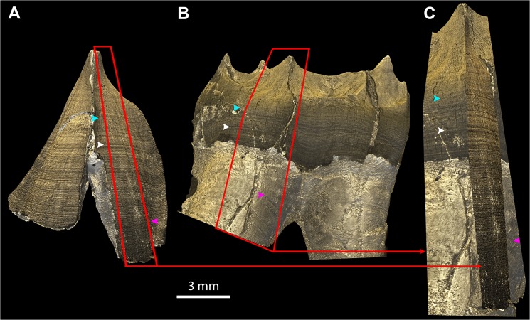
Fig 7. EDJ matching in multiple teeth of a single individual.
The 3D models of the EDJs of the LRC (A) and LRM1 (B) of KNM-KP34725 are matched in (C) by superimposing a portion of each EDJ (red frames and arrows) following the stress pattern as a barcode on the EDJ and root surface (colored arrowheads). This is done with Abode Photoshop by rotation, translation, isometric scaling, perspective and skewing of the fragment of EDJ of the canine onto the fragment of molar that is taken as a reference. (The two apical thirds of the roots of the molar were out of the field of view during scanning, thus the roots appear to be cut in an abrupt manner.) File name: Figure_7.tif.

