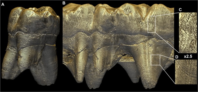
Fig 8. Unwrapped external surface of the StW151 ULM1.

Virtual unwrapping of the outer surface of the StW151 ULM1 obtained from the concatenation of a single pixel-wide frames saved during the complete rotation of the tooth around its long axis. On the left side of the unwrapped surface, the tooth is viewed from the buccal side, and the mesio-buccal and disto-buccal roots can be seen in the front, while the lingual root is visible in the back. The lingual view, at the right of the unwrapped tooth, shows the lingual root in the front, and the two buccal roots in the back. Since not all points are at the same distance from the center of rotation of the tooth, some parts of the tooth can be distorted: the furcation area of the buccal roots is stretched in the middle of the unrolled tooth. Perikymata, periradicular bands and hypoplasias are visible and can be tracked across the tooth. Images are not to scale. File name: Figure_8.tif.
