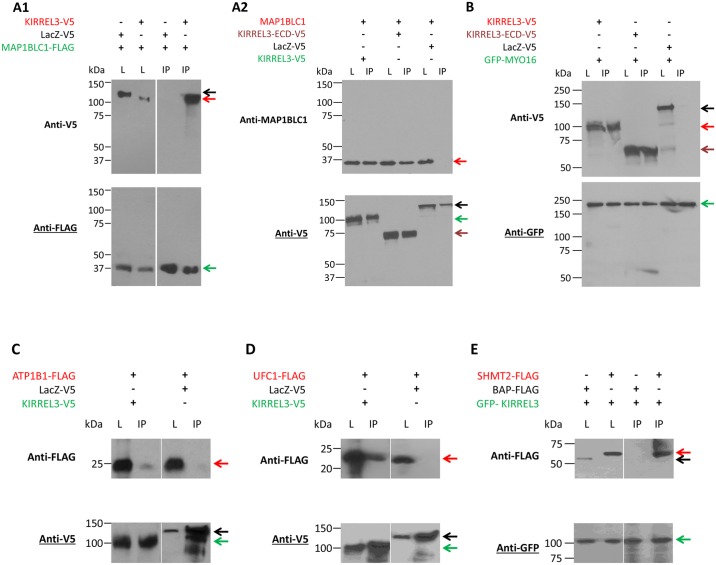
Fig 3. Western blot analysis of the Co-IP of KIRREL3-V5 with MAP1BLC1-FLAG (A1) and endogenous MAP1BLC1 (A2); KIRREL3-V5 with GFP-MYO16 (B), ATP1B1-FLAG (C), UFC1-FLAG (D), and GFP-KIRREL3 with SHMT2-FLAG (E).
Lysates from HEK293H cells overexpressing the indicated expression constructs were incubated with anti-FLAG antibody (A1), anti-V5 antibody (A2, C, and D), and anti-GFP antibody (B, E), and precipitated with magnetic beads. Lysates from N2a cells overexpressing KIRREL3-V5 expression construct was incubated with anti-V5 antibody, and precipitated with magnetic beads (A2). Immunoprecipitates (lane IP, Co-IP) were analyzed by western blotting as indicated with anti-V5, anti-MAP1BLC1, anti-FLAG, and anti-GFP antibodies. Expression of all proteins was also analyzed in total lysates (lane L). MAP1BLC1-FLAG (green arrow) immobilizes KIRREL3-V5 (A1, lane IP, red arrow), but not LacZ-V5 (black arrow). KIRREL3-V5 (green arrow) and KIRREL3-ECD-V5 (brown arrow), but not LacZ-V5 (black arrow) immobilize endogenous MAP1BLC1 (A2, lane IP, red arrow). GFP-MYO16 (green arrow) immobilizes KIRREL3-V5 (B, lane IP, red arrow), KIRREL3-ECD-V5 (B, lane IP, brown arrow) but not LacZ-V5 (B, lane IP, black arrow). (C) KIRREL3-V5 (green arrow) but not LacZ-V5 (black arrow) immobilizes ATP1B1-FLAG (red arrow). (D) KIRREL3-V5 (green arrow) but not LacZ-V5 (black arrow) immobilizes UFC1-FLAG (red arrow). (E) GFP-KIRREL3 (green arrow) immobilizes SHMT2-FLAG (red arrow) but not BAP-FLAG (black arrow).

