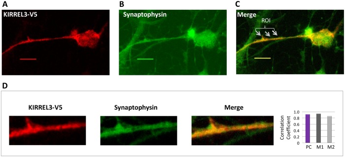
Fig 7. KIRREL3 colocalizes with the synaptic vesicles marker synaptophysin.
Rat PNCs overexpressing KIRREL3-V5 (red signal) (A) were immunostained with synaptophysin (green signal) (B). Yellow/orange signals (arrows in merged image) (C) in selected areas in a cell with KIRREL3-V5 suggested the colocalization of KIRREL3 with the synaptic vesicles. An enlarged overlay image and individual red and green channels for an area with ROI are shown (D). The degree of colocalization between the red and green signals was statistically analyzed (D) and expressed with Pearson’s correlation coefficient (PC) and Mander’s colocalization coefficients (M1 and M2). Bar, 20μm.

