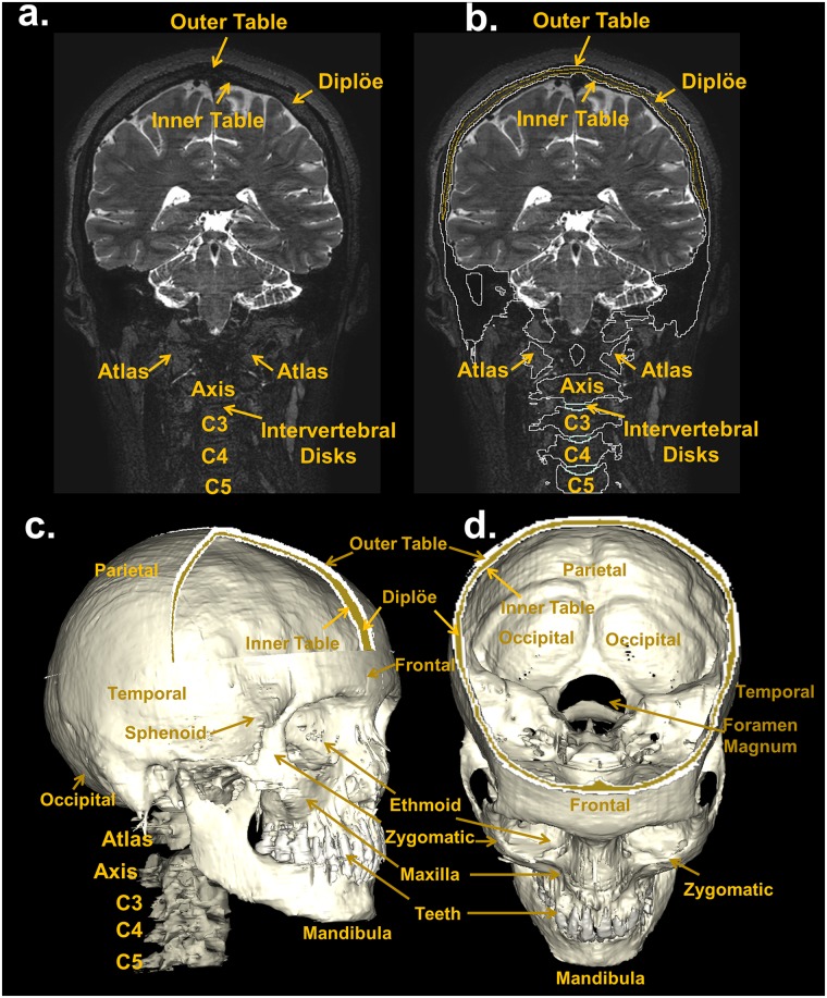
Fig 15. Segmentation of the bone.
Top: coronal view of a T2-weighted MRI slice (a) with the skull, vertebrae, and intervertebral disks outlined (b). The T2-weighted MRI, in which the intensity of the cancellous bone inside the diploë is enhanced compared to that of the cortical bones of the inner and outer tables made the subdivision of the skull into the main three layers, i.e., outer table (white outline), diploë (yellow outline), and inner table (white outline) possible. Bottom: 3D reconstruction of the skull, vertebrae, and intervertebral disks (c and d).

