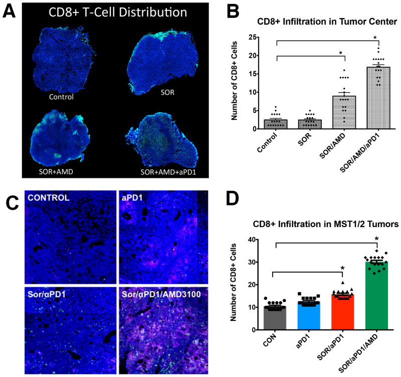
Figure 5. Anti-PD-1 treatment combined with sorafenib and AMD3100 increases intratumoral CD8+ T lymphocyte distribution in HCC.
A–D, The number of cytotoxic T lymphocytes infiltrating the tumor proper is increased only in the SOR+AMD3100+αPD1 treatment group. Representative confocal microscopy of immunofluorescence for CD8+ T lymphocytes (FITC, green) in HCC tissue sections (nuclei by DAPI, in blue) from HCA-1 grafted tumors (A) and representative regions of HCC tumors in the Mst GEM model. Red areas in the MST tumors indicate apoptotic regions of MST HCC tumors (staining for cleaved-caspase 3, Cy5 in red) (C). (B,D) Addition of anti-PD-1 antibody changes the distribution of CD8+ T lymphocytes in the tumors: Triple combination treatment results in significantly higher numbers of cytotoxic T lymphocytes in tumor center. Data are mean±SEM (n =5–6 per group) *P<0.05 vs. control.

