
Fig 6.
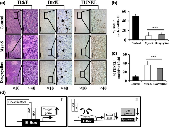
Histopathology of xenografts in nude mice and illustration of potential mechanism by Myc-5. (a) Tissue sample were analyzed qualitatively for morphological changes. Magnification, ×10 (scale bar = 200 μm); magnification, ×40 (scale bar = 50 μm). (b, c) Quantitative data of immunohistochemical analysis of BrdU and TUNEL positive staining in each group. Data in (b, c) are shown as the mean ± SD of three tumor samples from an individual mouse in each group. Statistical significance was calculated by Student's t-test. ***P < 0.001. (d) Schematic diagram of the mechanism by which pyrrole–imidazole polyamide inhibits MYC/MAX interaction to the E-box. (I) MYC:MAX dimer binds to E-box and activates MYC target gene expression. (II) Myc-5 occupied the E-box by binding, thereby inhibiting the MYC/MAX interaction to the E-box, causing further suppression of target gene expression.