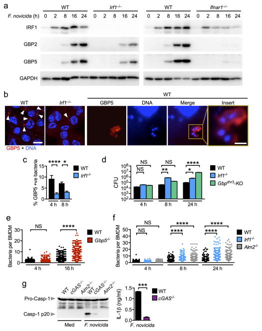
Figure 5. GBPs target bacteria to mediate killing during F. novicida infection.
(a) Induction of IRF1, GBP2 and GBP5 expression in unprimed BMDMs infected with F. novicida (MOI 50). (b) Immunofluorescence staining of GBP5 in unprimed BMDMs infected with F. novicida for 16 h. Scale bar, 10 μm; insert scale bar, 1 μm. (c) The percentage of bacteria colocalized with GBP5 was quantified. n=3,575 bacteria in WT BMDMs and n=3,620 bacteria in Irf1−/− BMDMs. (d) CFU recovered from WT, Irf1−/− and Gbpchr3-deleted BMDMs infected with F. novicida (MOI 100). (e) Quantification of the number of F. novicida bacteria per BMDMs using confocal microscopy. n=758 WT BMDMs and n=679 Gbp5−/− BMDMs. (f) Quantification of the number of F. novicida bacteria per BMDMs using confocal microscopy. n=715 WT BMDMs, n=710 Irf1−/− BMDMs, n=706 Aim2−/− BMDMs. (g) Caspase-1 activation and IL-1β release in unprimed BMDMs infected with F. novicida (MOI 100) for 20 h. Data from one experiment representative of two (d–f) or from three (a–c,g) independent experiments. *P < 0.05; **P < 0.005; ***P < 0.001; ****P < 0.0001; NS, not significant. Two-tailed t-test (c,g) and One-way ANOVA with a Dunnett’s multiple comparisons test (d) or with a Tukey’s multiple comparisons test (e,f).

