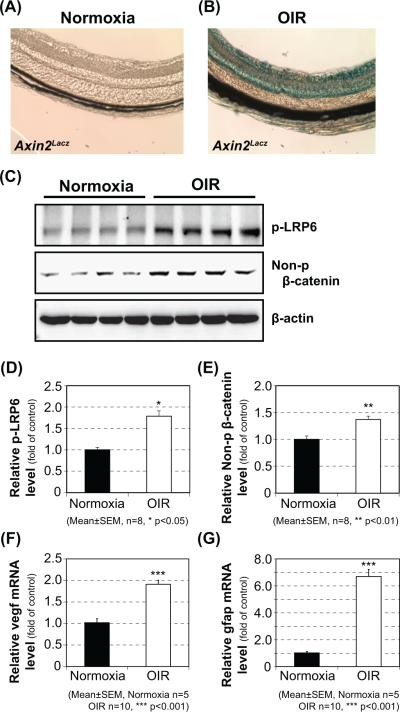
Figure 1. Activation of canonical Wnt signaling in the retina of OIR mice.
Wnt signaling activation was evaluated by X-gal staining of retinal sections of normoxia control (A) or OIR (B) Axin2Lacz mice at P16. Western blot analysis of phosphorylated-LRP6 (C, D) and non-phosphorylated-β-catenin (C, E) was performed using the retinas from the OIR C57BL/6J mice and normoxia control. The expression of Vegf-a (F), a major inflammatory factor, and Gfap (G), a commonly used stress marker, at the mRNA level in the retina of OIR mice was analyzed by qRT-PCR.

