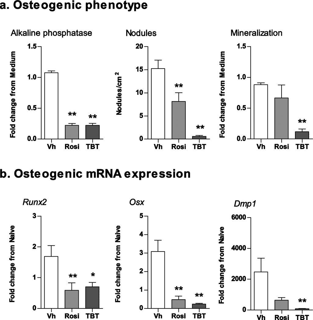
Figure 5. Rosiglitazone and TBT suppress osteogenesis in mouse BM-MSCs.
Primary bone marrow cultures were established from male C57BL/6J mice and treated with vehicle (Vh, DMSO), rosiglitazone (Rosi, 100 nM) or TBT (100 nM) in the presence of osteoinductive media for 7 (gene expression) or 11 days (osteogenesis assays). (A) Osteogenesis was assessed via alkaline phosphatase activity, alizarin staining and bone nodule counting. (B) mRNA expression was quantified by RT-qPCR. Data are presented as means ± SE (n = 5–8 independent bone marrow preparations). *p < 0.05, **p < 0.01 compared to Vh-treated cultures (ANOVA, Dunnett’s).

