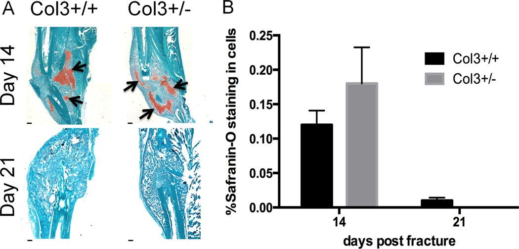
Figure 2. Histological analysis of fracture callus does not show differences in cartilage composition during healing.
A. Safranin-O staining in tibial fractures in wildtype and Col3 haploinsufficient mice (days 14 and 21 post fracture), revealing the presence of proteoglycan (arrows). By day 21, there is little to no cartilage present in the fracture callus. Scale bar= 400m. B. Quantification of percent cartilage found in fracture callus day 14 and 21 after fracture (n=4 or 5).

