
Table 1.
Magnetic Susceptibility of Common Biomolecules Encountered in SWI and QSM
| Materials | Volume susc.1 (SI, unitless) χv, ppm | Molar Susc.2 (CGS, cm3/mol, emu/mol) χm, ppm | Conditions and references | Areas of relevance |
|---|---|---|---|---|
| Water (H2O) | −9.035 | −12.98 | 20°C, Arrighini et al (47). | |
|
| ||||
| Hydroxyapatite (Ca2+) | −14.83 | Room temperature; measured by NMR; χv = −1.18 in CGS. Hopkins and Wehrli (111). | Vascular calcification, Tumor | |
|
| ||||
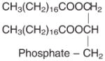
| Phospholipid | χ̄ = −9.68 | χ̄ = −5803 | 20°C, magneto-orientation method, phospholipid DPPC. Kawamura et al (63); phospholipid composes of stearic acids. | Myelination demyelination dysmyelination Multiple sclerosis |
|
χ|| = −10.43 | χ|| = −6303 | ||
| χ⊥ = −9.30 | χ⊥ = −560 | |||
| χa = −1.13 | χa = −683 | |||
|
| ||||
| Stearic acid CH3(CH2)16COOH | χ̄ = −10.03 | χ̄ = −218.4 | 20°C, Gouy method, powder stearic acid. ρ = 1.04 g/cm3, molar mass M = 284.4772 g/mol. Lonsdale 1939 (64). | |
| χ1 = −9.65 | χ1 = −210.0 | |||
| χ2 = −10.83 | χ2 = −235.7 | |||
| χ3 = −9.56 | χ3 = −208.2 | |||
| χa = −1.22 | χa = −26.6 | |||
|
| ||||
| Oxyhemoglobin (HbO2, Fe3+)4 | −893 | 20°C, Gouy method. Pauling and Coryell (90); Coryell et al (91). | BOLD fMRI Cerebral vascular diseases |
|
|
| ||||
| Deoxyhemoglobin (Hb, Fe2+) | 11,910 | 25°C, Gouy method, Taylor and Coryell (92). | ||
|
| ||||
| Methemoglobin (Hb+, Fe3+) | 14,000 | 24°C, Gouy method, in solution, per heme. Coryell et al (91). | Methemoglobinemia Sickle cell disease |
|
|
| ||||
| Hemosiderin (Fe2+ and Fe3+) | 4,810 | 22°C, Gouy method, thick suspension. Michaelis et el (94). | Hemorrhage Hemochromatosis |
|
|
| ||||
| Ferritin (Fe3+) | 6,132 | 22°C, Gouy method, in solution, per heme. Michaelis et el (94). | Brain iron stores Mitochondrial disease Neurodegeneration |
|
|
| ||||
| Ceruloplasmin (Cu2+) | 550 | 20°C, susceptometer balance, in solution, Ehrenberg et al (108). | Wilson’s disease | |
Shaded volume susceptibility values were converted from reported experimental molar susceptibility values (unshaded) in the relevant literature. Not all values were converted due to the unknown sample mass density and molar mass at the time of the reported experiments. The molar susceptibilities of phospholipid and oxyhemoglobin are much larger than that of water because of the presence of many more contributing chemical bonds within a molecule, see, e.g., Lonsdale (64).
Magnetic susceptibility values are reported in the literature based on several different definitions (volume, mass, and molar susceptibility) and units (SI, CGS, CGS emu). Significant variations exist due to variations in measurement techniques and sample preparations.
Conversion from molar to volume susceptibility follows where ρ is the mass density (g/cm 3) and M is the molar mass (g/mol). Conversion from CGS to SI units follows χv (SI) = 4πχv (CGS).
Mean susceptibility χ̄ = (χ1 + χ2 + χ3)/3; parallel susceptibility χ|| is the susceptibility along the long axis of the carbon chain; susceptibility anisotropy is χa = χ2 − (χ1 + χ3)/2 or χa = χ|| − χ⊥.
There are still debates on the iron state in oxyhemoglobin.