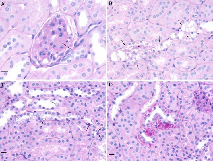
Fig. 6.
Histological appearance of db/db kidney sections stained with periodic acid-Schiff reagent (PAS) and counterstained with hematoxylin. All three of the db/db mouse groups contained the lesions noted in the images. a Segmental expansion of mesangium of a glomerulus (arrow). b Tubules with highly vacuolated epithelial cells (arrows) and PAS-positive material in lumen (asterisks). c Numerous variably enlarged clear to PAS-positive nuclei (so-called glycogenated nuclei) in tubular epithelial cells (arrows). d Accumulations of PAS-positive material in tubular epithelial cells. All scale bars represent 10 μm

