Fig. 2.
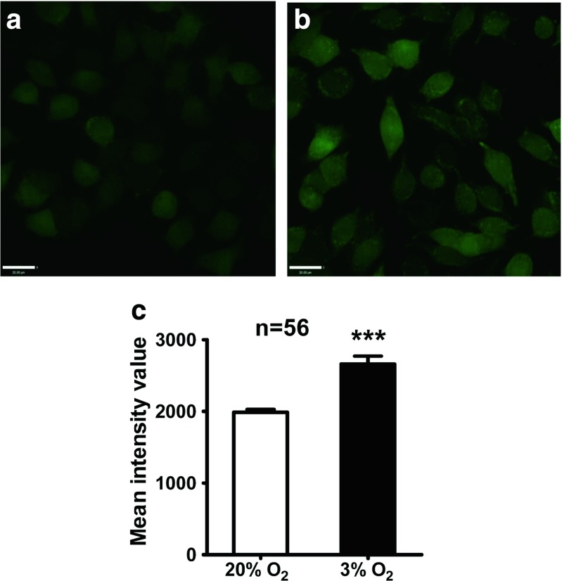
[Ca2+]i of PC12 cells in 20 and 3 % O2 as detected by calcium imaging. Hypoxic culture in 3 % O2 resulted in elevated [Ca2+]i in most PC12 cells (b), whereas calcium signals were relatively lower in PC12 cells cultured in 20 % O2 (a). The mean intensity value of basal [Ca2+]i in PC12 cells was higher in 3 % O2 compared with that in 20 % O2 (c). Scale bar = 20 μm; ***P < 0.001; “n” indicates the number of analyzed cells

