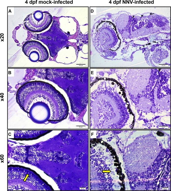
FIG 4.
Methacrylate-embedded sections of zebrafish larvae at 4 dpf stained with toluidine blue. (A to C) Control mock-infected larvae; (D to F) NNV-infected larvae showing marked neuropil vacuolation, as well as a relative paleness of neurons involving both the brain and retina. The most prominent injury appears to be in the photoreceptor layer (yellow arrows) of the retina, with apparent nearly total lysis of the photoreceptors. dpf, days postfertilization.

