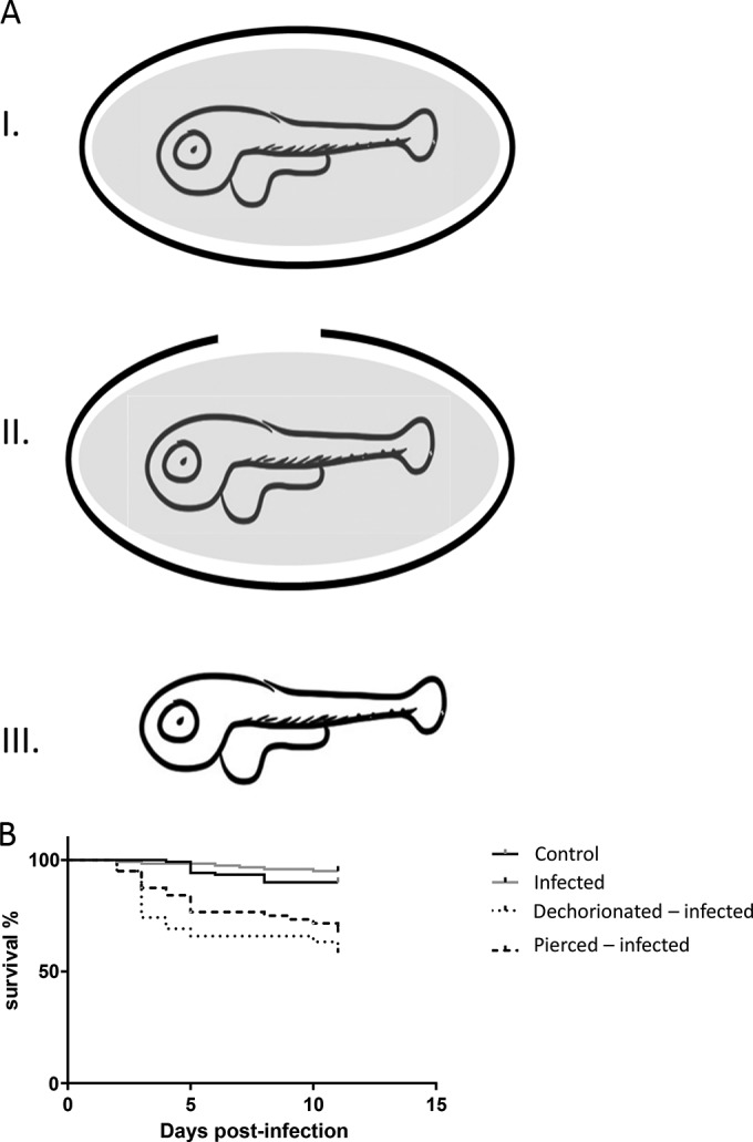
FIG 5.

Zebrafish larvae infected with NNV on day 2 postfertilization. (A) Schematic presentation of dechorionated larvae and pierced eggs. The schematics show eggs containing larvae (I), pierced larvae (II), and dechorionated larvae (III). The gray background represents perivitelline fluid. (B) Survival curves. The groups of eggs described in the legend to panel A were infected with NNV by bath immersion, and their survival rate was recorded. The data are representative of those from three independent experiments (n = 60 for each age group).
