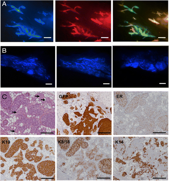
Figure 10.

Invasive tumor formation 6 and 8 weeks after intraductal injection of 4G-shp53-PI3K tumor cells. A. Mammary ducts dilated by human tumor cells at 6 weeks. B. Diffuse blue masses 8 weeks after intraductal injection caused by tumor cells invading the stroma. C. Mixed DCIS and invasive tumor at 8 weeks. H&E stain and immunohistochemistry show increased keratin 14 staining as cells migrate from the ducts into the stroma. Early squamous changes are marked by arrows. There are small difference in the structures in the serial sections in C because the structures gradually change as the sectioning proceeds through the paraffin block. Scale bars A 1 mm, B 2 mm, C 100 μm.
