
Figure 12.
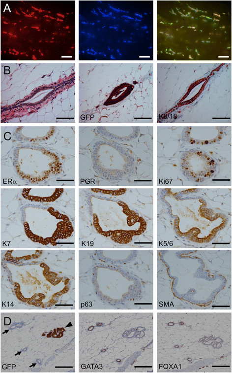
Replacement of the murine luminal layer 6 weeks after intraductal injection of 4G-shp53 cells. A. Stereomicrograph showing spread of human cells within the ducts. Alternating yellow and green areas in the right panel demonstrate independent engraftment of multiple clones. B&C. Histopathology (H&E stain) and immunohistochemistry show that the human cells have replaced the murine luminal layer with morphologically normal human cells. The human cells are larger, as seen at junctions between murine and human luminal cells. GFP, keratin 7 and keratin 19 staining specifically label the human cells. The cells are ERα+, PGR-, Ki67+ luminal cells. SMA staining shows that the myoepithelial layer is intact. D. GATA3 and FOXA1 are positive in murine ducts (marked by arrows) but negative in humanized ducts (marked by an arrowhead). Scale bars A 1 mm, B 100 μm, C 50 μm, D 200 μm.