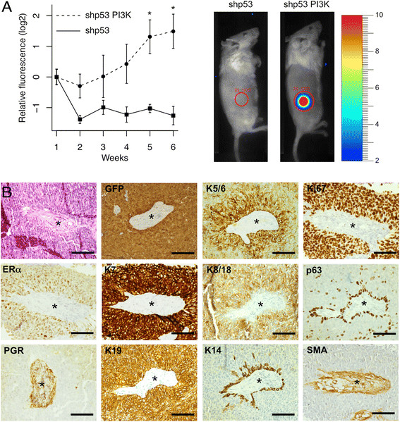
Figure 4.

Subcutaneous tumor formation requires activation of PI3K. A. Red fluorescence was used to measure subcutaneous tumor growth. Fluorescence in photons per second per cm2 per steradian was normalized to the starting value one week after injection. n = 6; *, p < 0.05. B. Histopathology (H&E stain) and immunohistochemistry show that the tumors were ERα+, Ki67+ adenocarcinomas. The central region in the sections contains an island of mouse stroma surrounded by human tumor cells (PGR gives a non-specific cytoplasmic stain in the stroma). Scale bars 100 μm.
