Figure 2.

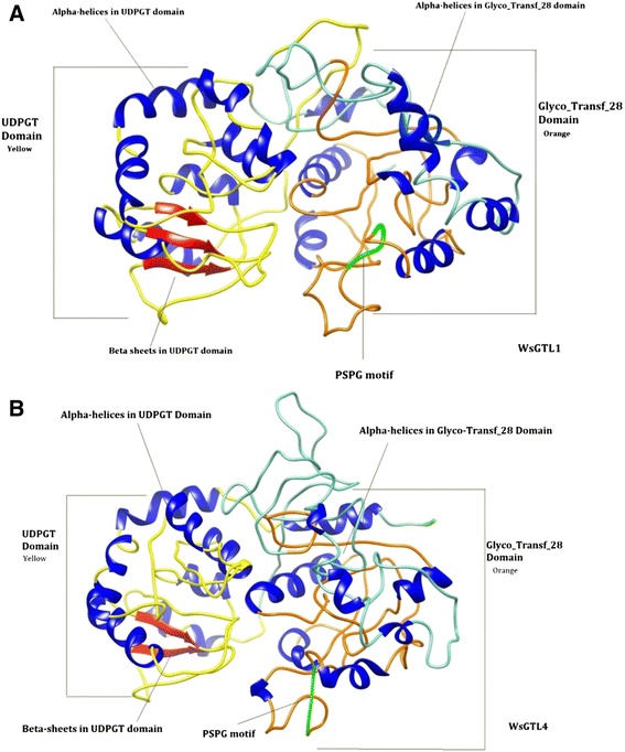
Ribbon diagrams of WsSGT proteins. (A) WsSGTL1 (B) WsSGTL4 showing Glyco_tranf_28 domain (orange), UDPGT domain (yellow), PSPG box (green), β-sheets (red) and α-helices (blue).

Ribbon diagrams of WsSGT proteins. (A) WsSGTL1 (B) WsSGTL4 showing Glyco_tranf_28 domain (orange), UDPGT domain (yellow), PSPG box (green), β-sheets (red) and α-helices (blue).