
Figure 3.
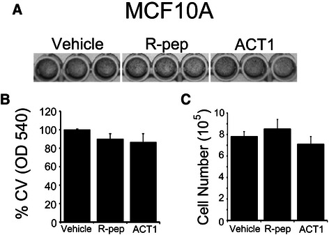
ACT1 does not alter proliferation of non-transformed MCF10A mammary epithelial cells. MCF10A cells were treated with vehicle, R-pep (200 μM), or ACT1 (200 μM) and assessed for (A) crystal violet staining density and (B) quantitated by OD540. (C) Cells were also assessed by cell counting. ± SEM; n = 6.