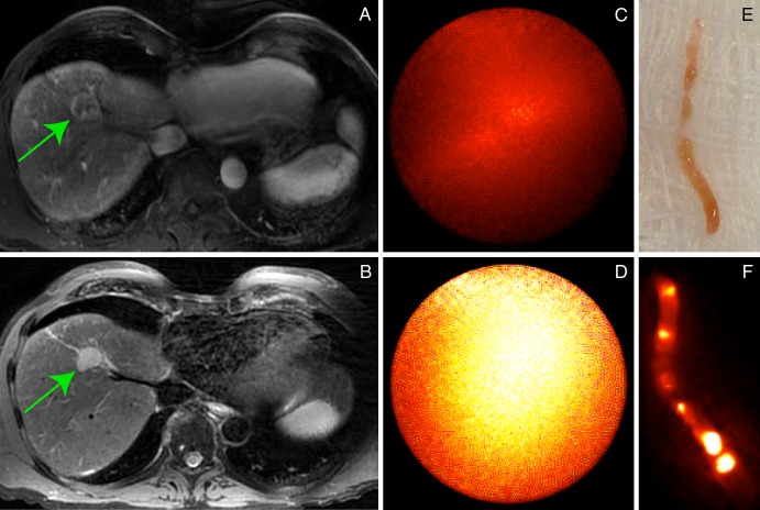
Figure 4:
Screening gadopentetate dimeglumine–enhanced MR images show, A, 21-mm enhancing lesion on a T1-weighted axial fat-suppressed image that demonstrates, B, intermediate-signal-intensity focal lesion (arrow) on a T2-weighted image in a 67-year-old man (patient 2) with HCV cirrhosis and prior history of renal cell carcinoma. Intraprocedural optical moIecular images demonstrate substantially increased ICG localization, D, in expected location of the lesion, C, relative to adjacent benign liver parenchyma during percutaneous biopsy. Surface reflectance imaging of, E, the core sample demonstrated, F, focal areas of increased ICG concentration. Final pathologic result was consistent with high-grade HCC.

