
Fig 1.
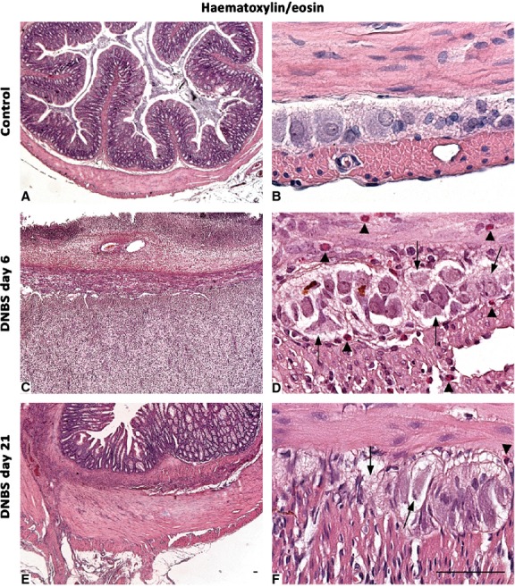
Histological appearance of haematoxylin/eosin-stained full-thickness colonic samples in control rats (A and B), or animals with DNBS-induced colitis at day 6 (C and D) and day 21 (E and F). The colonic wall of controls shows normal morphological features (A), with compact myenteric ganglia, which are plenty of neurons and glial cells (B). Colonic specimens from rats with colitis are damaged and thickened (C and E): myenteric ganglia appear to be vacuolized, with altered cells (arrows), and infiltrated by eosinophil granulocytes (D and F arrowheads), which are widely present also throughout the tunica muscularis; scale bars = 50 μm.