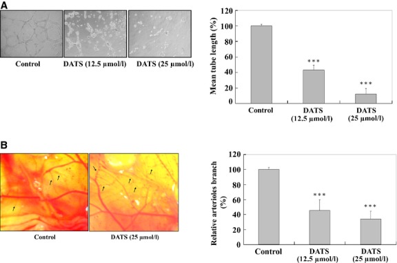
Fig 6.

DATS affected on VEGF-induced tube formation of HUVEC and an in vivoCAM assay. HUVEC (2 × 10⁵ cells) were incubated with or without 12.5 and 25 μM of DATS and then seeded in a 96-well culture plate pre-coated with Matrigel (BD Biosciences) and then were incubated for 24 hrs at 37°C in 5% CO2 atmosphere. After incubation, the cells morphology were evaluated by using a phase-contrast microscope and were photographed (200 × ; A). The quantitative data were determined using Image analysis software (B). On embryonic day 6 of fertilized White Leghorn chicken eggs, the developing CAM was separated from the shell by opening a small circular window at the broad end of the egg above the air sac. The eggs were incubated for two more days. Twenty-five μM DATS was prepared in PBS supplemented with 30 ng/ml of VEGF. On day 8, 20 μl was loaded onto 2-mm3 gelatin sponges as described in Materials and methods. Eggs were resealed and returned to the incubator. On day 10, images of CAM were captured digitally using an Olympus SZX9 stereomicroscope equipped with a Spot RT digital imaging system (C). The quantitative data indicated that the concentration of DATS was significantly different compared with control (D). ***P < 0.001 was considered significant.
