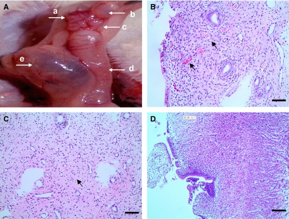
Fig 1.

Surgically induced rat model of EMs-affected oviduct by tissue autotransplantation in mesosalpinx, scale bar = 100 μm. (A) Macroscopic ectopic endometrial vesicle invades serous membrane of mesosalpinx and oviduct tissue in the experimental group, with diameter larger than 4 mm (black arrow), classified as grade III according to Quereda et al. (a) ovarian tissue; (b) ectopic endometrial vesicle; (c) oviduct tissue; (d) the left side of uterine horn; (e) local swelling and effusion after removal of the right side of uterine horn. (B) Hyperplasia and disturbance of capillaries (black arrows), indicated non-specific tissue reaction against the invasion of exogenous endometrial glands into oviduct wall. (C) Increased infiltration of lymphocytes and increased contents of fibre (black arrow), with abnormal hyperplasia of small vessels in EMs-affected oviduct wall, indicate chronic inflammation and interstitial fibrosis. (D) Normal oviduct tissue from the sham control.
