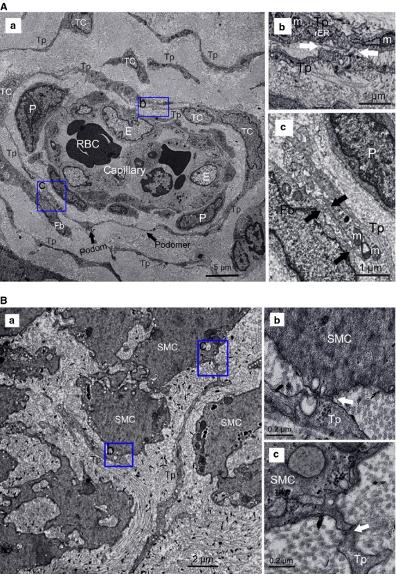
Fig 5.

Normal telocytes (TCs) with their telopodes (Tps), surrounding capillaries or scattered between smooth muscle bundles. (A) TCs around capillaries. (a) two or three layers of TCs formed a sheath around vascular endothelial cells (E) with their Tps, which composed of podom and podomer (black arrows), with pericytes (P) between them, Tps formed an almost complete circle to enwrap the capillaries. The organelles, such as mitochondria (M), rough endoplasmic reticulum (rER), cytoskeletal elements, can be observed. (b and c) Higher magnifications of the boxed areas; (b) TCs frequently established homocellular junctions with their Tps (white arrows); (c) heterocellular contacts between TCs and fibrocyte (Fb; black arrows). (B) TCs among smooth muscle cells (SMC). (a) Tp display close contact with SMC. (b and c) Higher magnifications of the boxed areas, show microvesicles visible in synaptic cleft (white arrows).
