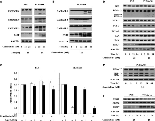
Fig 2.
Activation of PARP, caspases and other apoptosis mediators during HSP27-dependent gemcitabine-induced apoptosis. (A and B) Immunoblotting showing cleavage of PARP, CASPASE 3, CASPASE 8 and CASPASE 9 upon gemcitabine treatment in HSP27-overexpressing PL5/hu18 but not parental control cells in a dose-dependent (A) and time-dependent (B) manner. (C) Proliferation assays displaying abrogation of HSP27-dependent gemcitabine sensitivity through pre-incubation with the pan-caspase inhibitor Z-VAD-FMK at 20 μM for 3 hrs. Error bars represent SEM of at least three independent experiments. (D) Immunoblotting assessing expression changes of the indicated pro- or anti-apoptotic molecules in parental PL5 and PL5/hu18 cells upon treatment with gemcitabine at 25 nM at the indicated time-points. ß-ACTIN served as loading control. (E) Immunoblotting displaying time-dependent BIM expression changes in PL5/hu18 cells treated with gemcitabine at 25 nM. (F) Immunoblotting assessing expression changes of CHOP and GRP78 in parental PL5 and PL5/hu18 cells upon treatment with gemcitabine at 25 nM at the indicated time-points. ß-ACTIN served as loading control. Confirmatory HSP27 overexpression in PL5/hu18 cells is additionally depicted.

