
Fig 3.
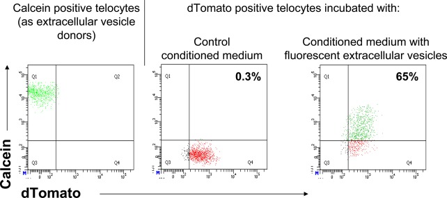
Mouse telocytes release and receive extracellular vesicles. Extracellular vesicle donor telocytes are negative for red fluorescent protein, dTomato (left panel), while extracellular vesicle receptor telocytes are positive for red fluorescence (middle and right panels). In left panel, extracellular vesicle donor telocytes are green fluorescent after incubation with calcein. Extracellular vesicle receptor telocytes are green fluorescent only after incubation with medium conditioned by calcein positive, extracellular vesicle donor telocytes (right panel), but not in case of conditioned medium derived from calcein negative cells (left panel).