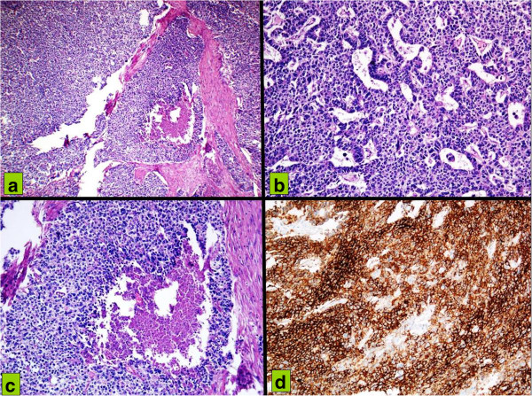
Figure 6.

Hematoxylin and eosin infiltration of gastric wall (a) by cells organized in a solid pattern with foci of necrosis (b) by neoplastic cells with pleomorphic nuclei and high nucleocytoplasmic ratio, with a trabecular and organoid pattern; (c) by tumor cells with vesicular nuclei, amphophilic cytoplasm, in a solid pattern of growth with central necrosis. (d) At immunohistochemistry neoplastic cells with high CD56 membrane positivity suspicious for neuroendocrine differentiation.
