
Figure 1.
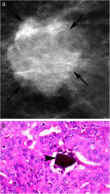
Mammogram and pathology report for HER-2 positive patient. a: Digital Mammogram (Mammomat Novation, Siemens Healthcare, Erlangen, Germany) of the left breast from a 40 year old woman with a HER-2 positive invasive ductal carcinoma, shows a malignant appearing mass (arrows) with numerous pleomorphic calcifications confined to the mass, BI-RADS 5. b: Hematoxyln and eosin stained section (400×) of poorly differentiated invasive ductal carcinoma with microcalcification (arrowhead).