
Figure 2. Various configurations of bacteria and biofilms in three different orthopedic patients.
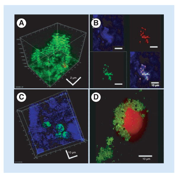
(A) Biofilm of live cocci (green) attached to a screw removed from a fixation device in a nonunion. The biofilm demonstrated classic 3D structure. (B) Patch of biofilm attached to periprosthetic tissue from a failed ankle arthroplasty. The upper left panel shows reflected light demonstrating the surface of the tissue (blue). The upper right panel shows a FISH ‘sau’ probe demonstrating Staphylococcus aureus bacteria (red). The lower left panel shows a FISH ‘Eub’ probe demonstrating all stained bacteria (green). The lower right panel shows an overlay demonstrating the S. aureus biofilm cluster attached to the tissue. (C) Periprosthetic tissue from the same patient as (B), showing bacteria that appear to be intracellular. (D) Intraoperative fluid from a patient with a failed elbow showing clumps of live cocci (green). The large red object is a nucleolus from a host cell that appears to have been ‘attacked’ and damaged by the cocci.