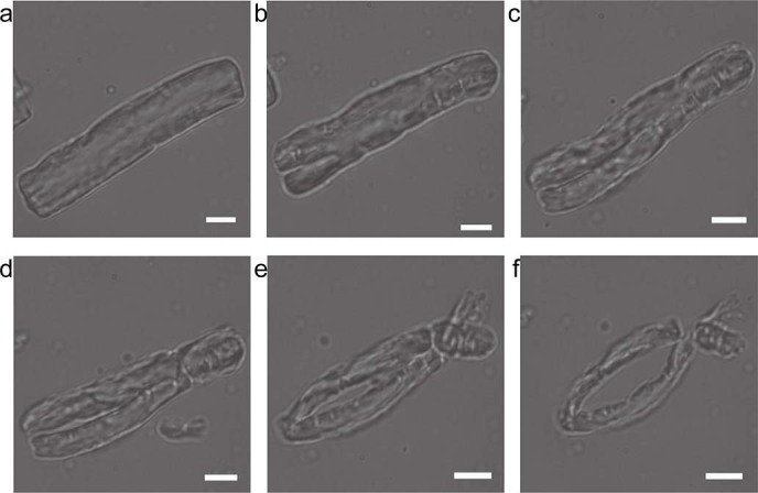
Figure 3. In situ observation of bovine elastin hydrolysis by pseudolysin using light microscopy.

Trace amount of bovine elastin was added to a drop of 1 mg/ml pseudolysin in 50 mM Tris-HCl buffer (pH 8.0) on a glass slide. The sample was observed and photographed with an inverted microscope (Olympus IX71, Japan) at room temperature at 5 min intervals. Magnification is ×960. Scale bars: 5 μm.
