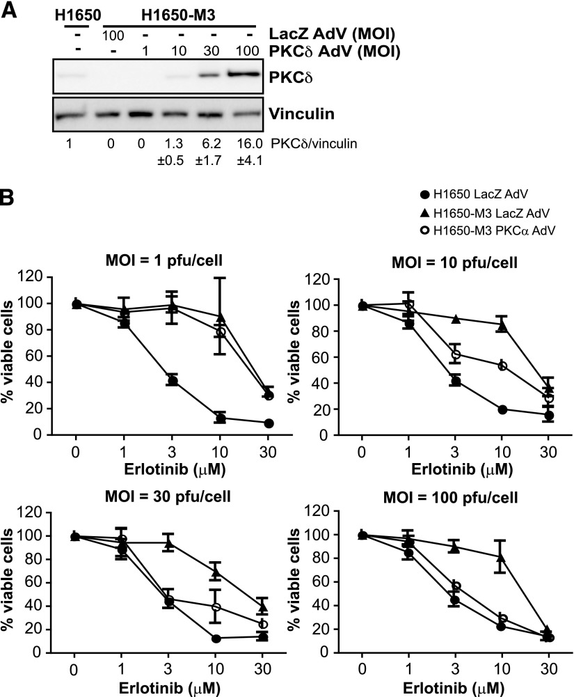
Fig. 3.
PKCδ alters the sensitivity of H1650-M3 cells to erlotinib. (A) H1650-M3 cells were infected with either PKCδ AdV or LacZ AdV at the indicated MOIs. Expression of PKCδ was determined using Western blot analysis. Densitometric analysis is shown as the mean ± S.D. (n = 3). (B) A viability assay using MTS was carried out 48 hours after infection. Data are expressed as the mean ± S.D. of triplicate samples. Similar results were observed in two additional experiments. pfu, plaque-forming unit.

