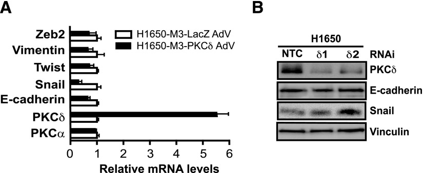
Fig. 6.
Genes involved in the mesenchymal phenotype are not regulated by PKCδ. (A) H1650-M3 cells were infected with either PKCδ AdV or LacZ AdV (MOI = 100 pfu/cell). After 96 hours, mRNA levels for various mesenchymal (vimentin, Snail, Twist, and Zeb2) or epithelial (E-cadherin) associated genes were measured by qPCR. Results are shown as the fold change relative to control (LacZ AdV-infected) H1650-M3 cells. Data were expressed as the mean ± S.D. of triplicate samples. (B) Parental H1650 cells were transfected with either PKCδ (PKCδ1 or PKCδ2) or NTC RNAi duplexes. Expression of PKCδ, E-cadherin, and Snail was analyzed by Western blotting 72 hours later. Similar results were observed in three independent experiments. NTC, nontarget control; pfu, plaque-forming unit.

