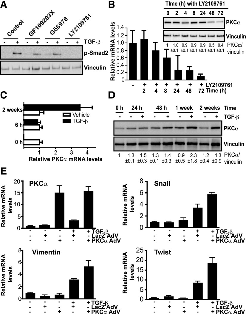
Fig. 7.
TGF-β signaling controls PKCα expression in erlotinib-resistant cells. (A) H1650-M3 cells were pretreated for 1 hour with either the pan-PKC inhibitor GF109203X (5 μM), the cPKC inhibitor Gö6976 (5 μM), the TGF-β receptor inhibitor LY2109761 (5 μM), or vehicle. Cells were then treated with TGF-β (20 ng/ml, 30 minutes) and phospho-Smad2 levels were determined by Western blot analysis. (B) H1650-M3 cells were treated with the TGF-β receptor inhibitor LY2109761 (5 μM) for the indicated times. PKCα mRNA and protein levels were determined by qPCR and Western blot analysis, respectively. Densitometric analysis is shown as the mean ± S.D. (n = 3). (C) PKCα mRNA levels in H1650 cells were measured 6 hours or 2 weeks after TGF-β treatment. (D) H1650 cells were treated with TGF-β (5 ng/ml) for 24 hours, 48 hours, 1 week, or 2 weeks. PKCα levels were determined by Western blot analysis. Densitometric analysis is shown as the mean ± S.D. (n = 3). (E) H1650 cells were infected with either PKCα AdV or LacZ AdV (MOI = 30 pfu/cell). Twenty-four hours after infection, cells were treated with TGF-β (5 ng/ml) for 1 week. mRNA levels for PKCα, Snail, vimentin, and Twist were measured using qPCR. In all cases, data were expressed as the mean ± S.D. of triplicate samples and experiments were reproduced at least three times. pfu, plaque-forming unit.

