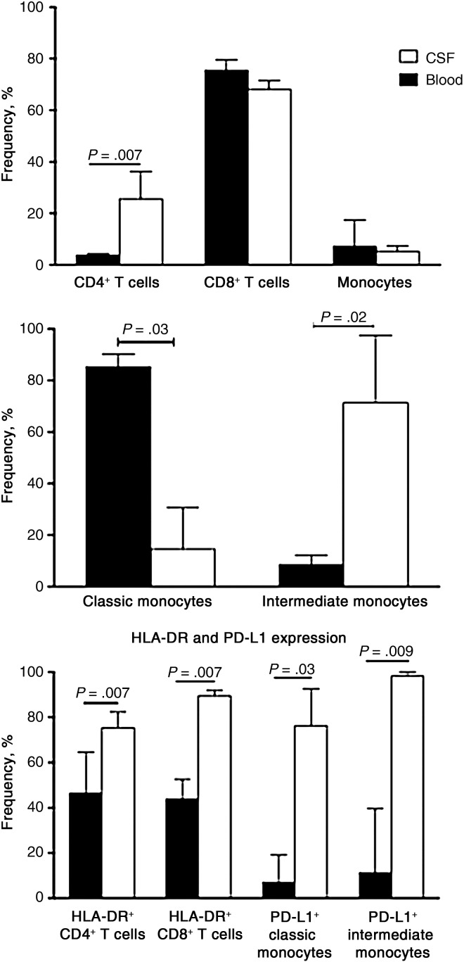
Figure 4.
Cell phenotype and activation in matched blood and cerebrospinal fluid (CSF) at diagnosis of cryptococcal meningitis with immune reconstitution inflammatory syndrome (CM-IRIS). Cell lineages in the CSF and peripheral blood compartments (top panel) and monocyte subsets (middle panel) are shown for 6 subjects at the diagnosis of CM-IRIS. The frequency of natural killer cells (not shown) was similar. Bottom panel shows HLA-DR expression on T cells and programmed death ligand 1 (PD-L1) expression on monocyte subsets.

