
Fig. 1.
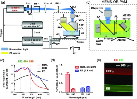
Optical-resolution PA microscopy (OR-PAM) systems. (a) Schematic of traditional OR-PAM system. AH, animal holder; BS, beam splitter; ConL, condenser lens; CorL, correction lens; M, mirror; MS, motorized stage; OL, objective lens; PH, pinhole; RAP, right angle prism; RhP, rhomboid prism; SOL, silicone oil layer; WT, water tank. (b) Schematic of fast-scanning MEMS-OR-PAM system. AC, aluminum coating; AL, acoustic lens. The MEMS-OR-PAM shares a similar light path outside the dashed box in (a). (c) Molar extinction spectra of oxy-hemoglobin (), deoxy-hemoglobin (HbR), and Evans blue (EB). While 560 and 595 nm were used for two-wavelength measurement on traditional OR-PAM system, 532 nm was used for dynamic measurement on MEMS-OR-PAM. (d) The average PA signals of blood- and 0.3% EB-filled plastic tubes at 560 and 595 nm. (e) The identification of the blood-filled (shown in red) and EB-filled (shown in green) tubes by comparing the PA signals at two wavelengths.