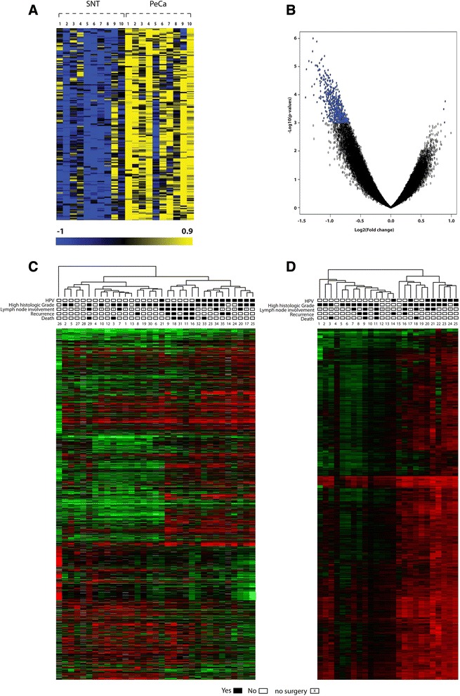
Figure 1.

Supervised and unsupervised analysis of gene expression and methylation profiles. (A) Heat map showing 171 significantly hypermethylated probes in the paired analysis of 10 PeCa and SNT samples (P value ≤0.001 and FDR ≤0.05). (B) Volcano plot of the methylated regions: left-sided deviation indicates that all hypermethylated probes were tumor related. (C) Unsupervised analysis showing dendogram and heat map of altered transcripts from the gene expression analysis in PeCa. (D) Unsupervised analysis showing dendogram and heat map of altered probes from the methylation analysis in 25 PeCa samples. Rectangles show clinical characteristics of patients with PeCa. Numbers below rectangles represent samples.
