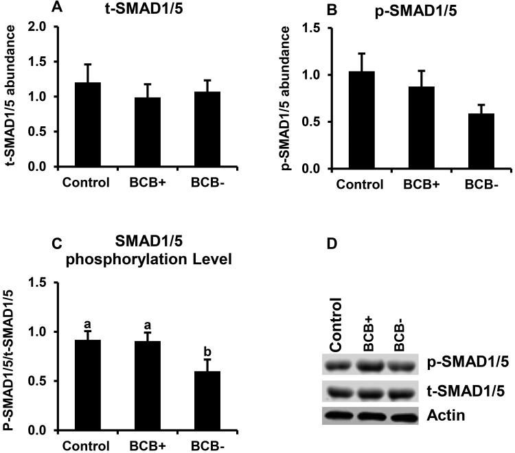
Figure 6.
SMAD1/5 phosphorylation in BCB-screened, GV-stage oocytes. Samples of control, BCB+ and BCB− GV stage oocytes (n=6 replicates of 20 oocytes/group) were subjected to Western blot analysis of total (t)-SMAD1/5 (A), phosphorylated (p)-SMAD1/5 (B). Expression levels were normalized relative to abundance of endogenous control (actin). Phosphorylation level (C) was expressed as p-SMAD1/5/t-SMAD1/5 Data are shown as mean ± standard error. Values with different superscripts across treatments indicate significant differences (P < 0.05). Representative Western blot (D).

