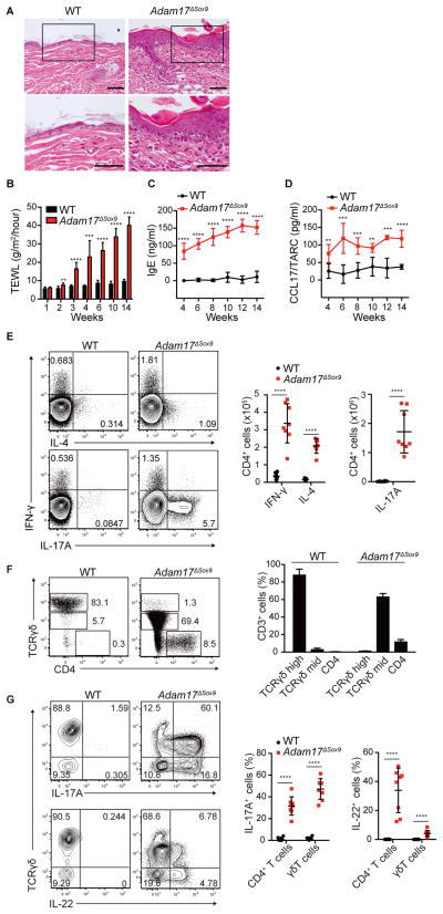
Figure 1. Eczematous inflammation in ADAM17-deficient skin.
(A) Histopathology of skin biopsy from WT and Adam17fl/flSox9-Cre (Adam17ΔSox9) mice. Scale bars, 50 μm. (B–D) TEWL, serum IgE and serum CCL17/TARC values in WT and Adam17ΔSox9 mice (N=8). (E) Flow cytometry analysis of IFN-γ, IL-4 and IL-17A production in CD4+ cells from lymph nodes, and (F, G) IL-17A and IL-22 production by CD3+ cells in epidermis of WT and Adam17ΔSox9 mice (N=8). TCRγδhigh cells represent dendritic epidermal T cells, and TCRγδmid cells represent γδ T cells that have infiltrated epidermis from the dermis. Data in B–G are shown as mean ± SD. *P<0.05, **P<0.01, ***P<0.001, ****P<0.0001 as determined by Student’s t test. See also Figures S1.

