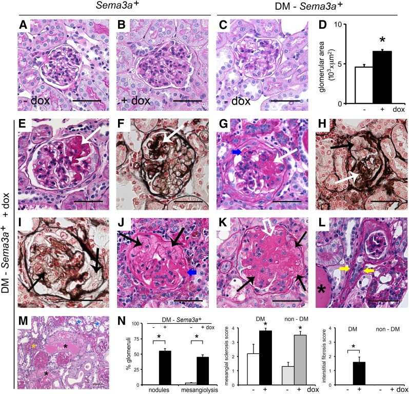
Figure 3.
Sema3a+ gain of function in diabetic mice causes advanced diabetic nephropathy. Periodic acid Schiff (PAS) stain of nondiabetic kidneys (Sema3a+) shows normal histology in control kidney (-dox) (A), whereas a kidney from a mouse receiving doxycycline (+dox) shows mesangial expansion (B). C: PAS staining of a biopsy sample from a control diabetic kidney (diabetes mellitus [DM]-Sema3a+ -dox) shows mild mesangial expansion. D: Quantification of glomerular area indicates that Sema3a+ gain of function in diabetic mice induces glomerulomegaly (DM-Sema3a+ +dox vs. DM-Sema3a+ -dox; n = 4 kidneys each, 34 ± 2 glomeruli/kidney). E–M: PAS and Jones’ silver stains of Sema3a+ gain-of-function diabetic kidneys (DM-Sema3a+ +dox) show nodular Kimmelstiel-Wilson-like glomerulosclerosis (white arrows), mesangiolysis (black arrows), diffuse glomerulosclerosis (white open arrow), fibrin caps (blue arrows), arteriolar hyalinosis (yellow arrows), foam cells (blue asterisks), protein casts (black asterisks), and interstitial infiltrates (yellow asterisk). N: Quantification of glomerular nodules and mesangiolysis, shown as a percentage of glomeruli with nodules or mesangiolysis per kidney in diabetic Sema3a+ gain-of-function (+dox) vs. diabetic control mice (-dox) (134 ± 6 and 121 ± 5 glomeruli/kidney were counted in n = 5 and n = 4 kidneys, respectively; P < 0.05). Semiquantitative pathology score shows significantly increased mesangial sclerosis in all Sema3a+ gain-of-function kidneys, whereas interstitial fibrosis occurs exclusively in Sema3a+ gain-of-function diabetic mice (black bars). Scale bars = 50 μm (A–C, E–L). *P < 0.05, Sema3a+ gain-of-function vs. diabetic control mice.

