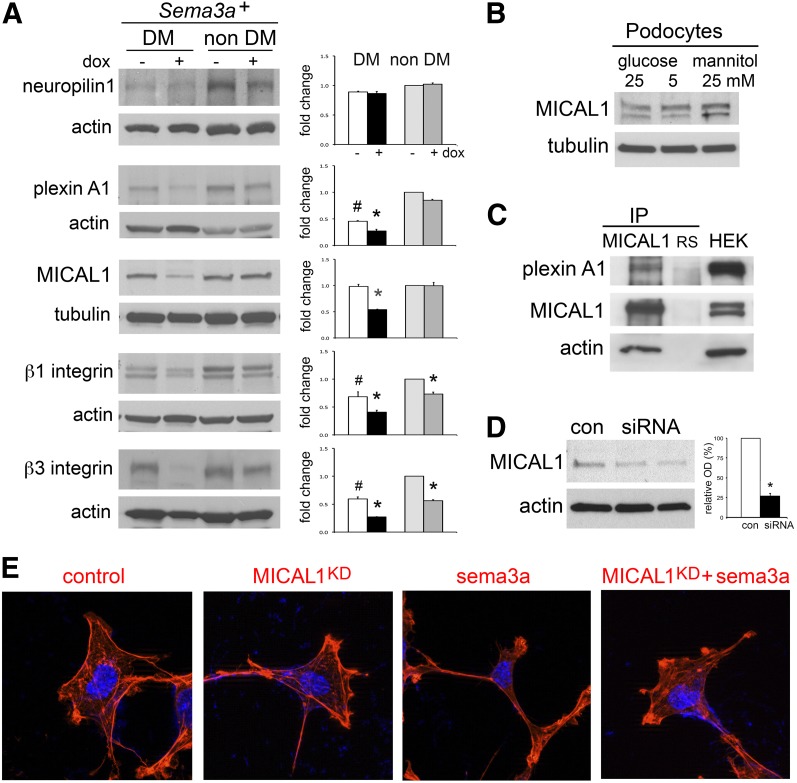
Figure 7.
Sema3a signals in podocytes are mediated by MICAL1. A: Western blots show that the sema3a signaling pathway is expressed in the kidney. PlexinA1, MICAL1, and β3 integrin are downregulated in Sema3a+ gain-of-function diabetic (DM) mice (black bar). Quantitation by densitometry is shown in adjacent bar graphs. Data are expressed as mean ± SEM from three or more independent experiments. B: MICAL1 is expressed in cultured podocytes and is not altered by 4-h exposure to high glucose. C: Coimmunoprecipitation (IP) demonstrates an endogenous plexinA1–MICAL1 interaction in podocytes; actin coprecipitates with the plexinA1–MICAL1 complex. Rabbit serum (RS) and whole-cell lysate from HEK cells transiently transfected with full-length MICAL1 (HEK) were used as controls. D: Immunoblot shows MICAL1 knockdown of ∼75% by siRNA, confirmed by densitometric analysis. E: MICAL1 knockdown (KD) prevents sema3a-induced podocyte contraction and F-actin collapse, assessed by rhodamine-phalloidin staining. Data from three or more independent experiments are shown. *P < 0.05 vs. corresponding control. #P < 0.05 vs. nondiabetic control. OD, optical density.

