Figure 9.
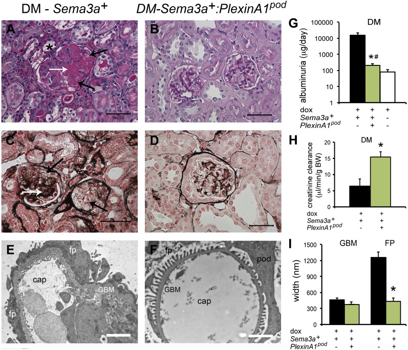
Deletion of podocyte plexinA1 attenuates diabetic nephropathy in mice. A–D: Periodic acid Schiff and Jones’ silver stains show severe diabetic (DM) nodular glomerulosclerosis in Sema3a+ gain-of function kidneys (A and C) and mild mesangial expansion and otherwise normal histology in diabetic Sema3a+:plexinA1pod kidneys (B and D). A: *, foam cell; A and C: white arrows, nodule; black arrows, mesangiolysis. Scale bars = 50 μm. TEM shows complete foot process (FP) effacement, thickened GBM, and endothelial swelling in Sema3a+ gain-of-function diabetic glomeruli (E), whereas TEM of Sema3a+:plexinA1pod mice shows very mild GBM thickening and virtually no FP effacement (F), as confirmed by morphometric analysis (n = 4 per group; I). Scale bars = 2 μm. G and H: Deletion of podocyte plexinA1 in diabetic mice results in mild albuminuria and normal creatinine clearance (green bars), similar to that in wild-type diabetic mice (white bar), whereas Sema3a+ gain of function causes massive albuminuria and renal insufficiency (black bars). Black bars are diabetic sema3a gain-of-function (+dox); green bars are diabetic plexinA1 knockout + sema3a gain-of-function (+dox); white bars are diabetic controls (-dox). *P < 0.05 vs. Sema3a+ gain of function. #P < 0.05 vs. wild-type diabetic mice. BW, body weight; cap, capillary lumen; dox, doxycycline; pod, podocyte.

