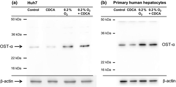
Fig 5.

Hypoxia treatment for 18 h increases OSTα protein levels in Huh7 cells and PHH. Cells were treated with 50 μM CDCA or DMSO alone and were either exposed to hypoxia or kept under normoxic conditions for 18 h. Total protein from whole Huh7 cell extracts (30 μg) (a) or 15 μg from whole cell extracts isolated from PHH (b) were run on an SDS-PAGE gel and immunoblotted. An anti-OSTα antibody was used to probe the membrane, followed by stripping and probing with β-actin antibody. Exposure of the cells to hypoxia-induced protein expression in both Huh7 (three-fold) and PHH (two-fold).
